How traits pass from one generation to the next: the cell and its chromosomes, mitosis and meiosis, Mendel’s laws, and the ways genes interact to shape the animals we breed. This is the entry point to the ASAP-Bio breeding track.
Genetics is the science of heredity and variation: how characteristics are transmitted from parents to offspring, and why individuals differ. For animal scientists it is the foundation of breeding, because we can only improve what is inherited. The field has several branches, including transmission (Mendelian) genetics, molecular genetics, population genetics and quantitative genetics, the last two being the bridge into animal breeding.
A small, exact vocabulary carries the whole subject. A gene is a unit of heredity occupying a fixed position, its locus, on a chromosome. Alternative forms of a gene are alleles. An animal’s genetic make-up is its genotype; the observable result is its phenotype. When the two alleles at a locus are identical the animal is homozygous; when they differ it is heterozygous. An allele whose effect is seen in the heterozygote is dominant; one whose effect is masked is recessive.
These terms are not pedantry; they are the difference between guessing and predicting. A breeder who knows an animal’s genotype can predict what it will transmit, whereas the phenotype alone can be misleading because a heterozygous carrier looks identical to a homozygous dominant. Coat colour makes this concrete: a black animal may be BB or Bb, and only breeding records or a DNA test separate the two. The same logic governs genetic defects: many congenital and lethal abnormalities are recessive, so healthy-looking heterozygous carriers silently pass them on until two carriers are mated. Identifying and avoiding carriers of bad genes is therefore one of the first practical uses of genetics in a breeding herd.
Context on how variant catalogues are built and represented.
Genes are carried on chromosomes. Body (somatic) cells are diploid (2n): chromosomes come in homologous pairs, one from each parent. Gametes are haploid (n), carrying a single set. Cattle, for example, have 2n = 60; sheep 2n = 54; goats 2n = 60. Two kinds of division maintain and reshuffle this material.
Mitosis divides a somatic cell into two genetically identical daughter cells, used for growth and tissue repair. After DNA replication, the cell passes through prophase, metaphase, anaphase and telophase: chromosomes condense, line up on the cell’s equator, sister chromatids separate, and two identical nuclei form. The chromosome number is conserved (2n → 2n).
Meiosis produces gametes and halves the chromosome number (2n → n) over two successive divisions. It is the source of genetic variation through two mechanisms. In prophase I, homologous chromosomes pair and exchange segments by crossing over (recombination). At metaphase I / anaphase I, the homologous pairs line up and separate independently of one another, so maternal and paternal chromosomes are dealt into gametes in new combinations. The result is four haploid cells, each genetically unique.
Both divisions are preceded by interphase, in which the cell grows and, in the S phase, copies all its DNA so each chromosome consists of two identical sister chromatids joined at a centromere. Mitosis then separates those sisters once, conserving the number; meiosis separates homologous chromosomes in the first division and sisters only in the second division, which is why one starting cell yields four haploid products rather than two. The biological pay-offs differ accordingly: mitosis underlies growth, wound healing and the faithful replacement of worn tissues, and errors in its control are the basis of tumours, whereas meiosis exists precisely to create variation and to halve the genome so that fertilisation can restore it. For a breeder, meiosis is the step that shuffles a superior parent’s genes into many different gametes, which is why even an outstanding sire produces offspring of varying merit.
At the chromosomal level each species has a characteristic karyotype, the number and shape of its chromosomes: cattle have 2n = 60, the pig and the cat 2n = 38, and the chicken 2n = 78 (with ZZ males and ZW females, the reverse of the mammalian XX/XY system). Chromosome aberrations matter directly to breeders. Robertsonian translocations (centric fusions of two chromosomes), of which the 1/29 fusion in cattle is the classic example, leave the carrier outwardly normal but produce unbalanced gametes, lowering fertility and causing early embryonic loss; numerical errors such as trisomy are usually lethal. Karyotyping and chromosome-painting techniques are therefore used to screen breeding animals, especially valuable sires, for hidden structural defects before they are used widely.
Gametogenesis is the formation of gametes by meiosis. In the male, spermatogenesis in the testis turns one diploid spermatogonium into four functional spermatozoa. In the female, oogenesis in the ovary yields a single large ovum per meiosis (the other products become polar bodies), because the egg retains almost all the cytoplasm and nutrients.
At fertilisation, a haploid sperm (n) fuses with a haploid egg (n) to form a diploid zygote (2n), restoring the species chromosome number and uniting the genetic contributions of both parents. The offspring’s genotype is therefore one new sample from each parent’s reshuffled genome, which is exactly why selection of parents changes the next generation.
The two processes also differ sharply in timing and number. Spermatogenesis runs continuously from puberty, producing sperm in enormous numbers throughout the male’s life, which is what makes one sire able to inseminate many females. Oogenesis is far more economical: females are born with a fixed stock of arrested oocytes, and typically only one (or a few) completes maturation and ovulation in each reproductive cycle. At fertilisation a single sperm penetrates the ovum and the egg immediately raises a block to further sperm (preventing polyspermy), after which the two haploid nuclei fuse. The sperm also carries the sex chromosome (X or Y in mammals) that determines the offspring’s sex, so it is the male gamete that decides whether a calf is male or female.
Gregor Mendel (1865) deduced the rules of single-gene inheritance from pea crosses, and they apply directly to animals. The law of segregation: the two alleles at a locus separate during gamete formation, so each gamete carries only one. The law of independent assortment: alleles of different genes are distributed to gametes independently (for genes on different chromosomes).
A cross of two heterozygotes for one gene, Bb × Bb, gives genotypes 1 BB : 2 Bb : 1 bb, and with full dominance a 3:1 phenotype ratio. A cross of two double heterozygotes, BbEe × BbEe, gives the classic 9:3:3:1 phenotype ratio because the two genes assort independently. A test cross (mating a dominant-looking animal to a recessive homozygote, bb) reveals the unknown genotype: any recessive offspring prove the parent was a carrier (Bb).
Mendel succeeded where others had failed because he counted offspring over many crosses and reasoned that inheritance is particulate, alleles stay intact and segregate, rather than blending. The dihybrid case shows the power of his second law. Crossing two animals heterozygous for coat colour and a second gene, BbEe × BbEe, each parent makes four equally frequent gamete types (BE, Be, bE, be); combining them in a 4×4 Punnett square gives sixteen boxes that collapse to the famous 9:3:3:1 phenotype ratio when both genes show simple dominance. The test cross is the practical tool that falls out of this theory: mating a dominant-looking animal to a recessive (bb) tester converts hidden genotypes into visible offspring ratios, a 1:1 split betrays a heterozygote, while all-dominant offspring point to a homozygote.
Many traits depart from clean dominance. Under incomplete dominance the heterozygote is intermediate (a red × white cross giving roan-like blends). Under codominance both alleles are fully expressed in the heterozygote (the AB blood group; many protein and DNA markers). A locus may have multiple alleles in the population even though any one animal carries only two, as with coat-colour series and blood-group systems. Some alleles are lethal, removing a genotypic class and distorting ratios (for example a 2:1 ratio when the homozygote dies).
Epistasis is interaction between genes: one locus masks or modifies another. It changes the dihybrid 9:3:3:1 into recognisable ratios, including 9:3:4 (recessive epistasis), 13:3 (dominant suppression), 9:7 (duplicate recessive / complementary genes), 15:1 (duplicate dominant) and 12:3:1 (dominant epistasis). Two further ideas matter for breeders: pleiotropy, where one gene affects several traits, and the fact that the environment can control expression (temperature-sensitive coat colour; nutrition and growth genes). These complications are exactly why most economically important traits are treated quantitatively, the subject of the next course.
The course materials give memorable animal examples of each departure. In mice, coat colour shows recessive epistasis: an agouti × agouti cross can segregate agouti : black : albino in a 9:3:4 ratio because a homozygous-recessive colour gene leaves the animal albino regardless of the agouti locus. In dogs, a dominant inhibitor gene (I) suppresses black pigment, giving the dominant-epistasis ratios (such as 13:3) where one gene overrides another. The Himalayan / Siamese pattern shows the environment controlling expression directly: the pigment enzyme is temperature-sensitive, so the same genotype is dark on the cool extremities and pale on the warm body, white above about 30°C. Multiple alleles appear as a series, the rabbit coat-colour series (full colour, chinchilla, Himalayan, albino) being the classic case, even though any one rabbit carries only two of them. And a lethal allele removes a genotypic class, turning an expected 1:2:1 into a 2:1 ratio among survivors. Each pattern is still Mendelian at heart; the ratios simply reveal genes acting together.
Coat colour gives a concrete biochemical picture of how these genes interact. Pigment is melanin in two forms, dark eumelanin and reddish-yellow phaeomelanin, synthesised in melanocytes from the amino acid tyrosine by the enzyme tyrosinase. The Extension (E) locus encodes the MSH receptor (MC1R) that drives production toward eumelanin, while the Agouti (A) locus encodes a signalling protein that blocks it and switches production toward phaeomelanin; further loci (the B and C pigment enzymes and the D dilution locus, which affects how pigment granules disperse) modify the result. Epistasis among them is mechanistic, not abstract: a non-functional C locus (albinism) blocks tyrosinase and masks every other colour gene, which is precisely why such crosses collapse to modified ratios. The classical colour genes thus become a worked example of how genotype maps, enzyme by enzyme, to phenotype.
Coat colour and the blood groups obey single-gene rules, but milk yield, growth rate and fertility do not fall into neat categories. They are polygenic, controlled by many genes of small effect, and strongly influenced by the environment, so they vary continuously. The Mendelian logic you have learned still holds at each locus; it simply sums over many loci. Capturing that sum statistically, through means, variances, heritability and breeding values, is the job of quantitative genetics and animal breeding.
The bridge is the idea of many loci summing together. If one gene gives a clean 3:1 split, a handful of genes acting on the same trait produce several overlapping classes, and dozens of genes, each adding a small plus-or-minus, blur into a smooth bell-shaped normal distribution of phenotypes. Add environmental variation on top and the categories vanish entirely: we can no longer label an animal “black or red” but must measure it (litres of milk, grams per day) and describe the population with a mean and a variance. Stretches of chromosome carrying genes that influence such traits are called quantitative trait loci (QTL), and modern genomics can map them, but the breeder rarely needs to know the individual genes. What matters is how much of the variation is heritable and transmissible, which is exactly the statistical machinery, heritability, breeding values and selection, that the next course builds.
Single-locus rules also scale up to whole populations. If an allele has frequency p, random mating distributes the genotypes in the Hardy-Weinberg proportions p² : 2pq : q², and a simple chi-square test of observed against expected numbers reveals whether a population is in equilibrium or is being disturbed by selection, migration, drift or non-random mating. Allele frequencies are estimated directly by the gene-counting method, and following how they shift , under selection against a recessive, heterozygote advantage (overdominance), or mutation-selection balance , is the quantitative heart of population genetics. The downloadable problem sets in this theme work these calculations through in full.
To see how this genetics underpins the conservation and sustainable use of livestock diversity, take the FAO course on Plant and Animal Genetic Resources (SDG indicators 2.5.1 & 2.5.2), and on managing genetic variation, Pre-breeding: Creating and Managing Variation.
FAO · Plant & Animal Genetic Resources → FAO · Pre-breeding →
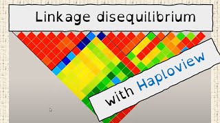
Genomic selection rests entirely on linkage disequilibrium between markers and causal variants. These show LD as a measurable, visualisable quantity.